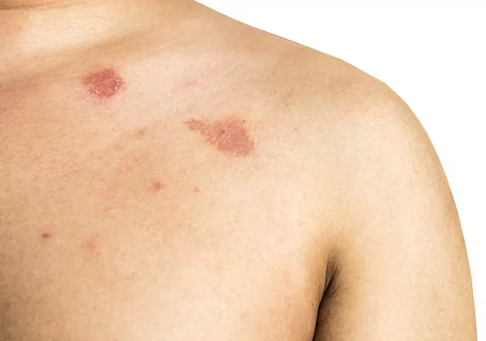
Tinea pedis, commonly known as athlete’s foot, is a dermatophyte fungal infection affecting the skin of the feet. This condition is widespread, affecting individuals of all ages worldwide. While the symptoms of tinea pedis are often uncomfortable and sometimes debilitating, understanding the causative agent behind this condition is crucial for effective treatment and prevention strategies. In this article, we delve into the world of fungi to unveil the culprit responsible for tinea pedis.
The Fungus Behind Tinea Pedis: Trichophyton rubrum
Among the various fungal species implicated in causing tinea pedis, Trichophyton rubrum stands out as the predominant culprit. T. rubrum belongs to the group of dermatophytes, which are fungi capable of invading and thriving on the keratinized tissues of the skin, hair, and nails. This species accounts for a significant proportion of dermatophyte infections globally, including tinea pedis.
Understanding Trichophyton rubrum: Morphology and Characteristics
Trichophyton rubrum is a filamentous fungus characterized by its septate hyphae, which form a network known as mycelium. Under the microscope, T. rubrum appears as branching hyphae with conidia, the asexual spores responsible for the spread of the fungus. These conidia are often produced in chains, giving the fungus a distinctive appearance.
In addition to its microscopic features, T. rubrum colonies grown in culture exhibit specific macroscopic characteristics. These colonies typically have a powdery texture and a pink to red coloration, although variations may occur depending on environmental conditions and the specific strain of the fungus.
Pathogenesis of Tinea Pedis
The pathogenesis of tinea pedis involves a complex interplay between the fungus, host factors, and environmental conditions. Trichophyton rubrum thrives in warm, moist environments, making the interdigital spaces of the feet an ideal habitat. Upon encountering favorable conditions, T. rubrum adheres to the keratinized tissues of the skin and begins to proliferate.
The fungus secretes enzymes, such as keratinases, which enable it to break down keratin, the predominant protein in the outer layers of the skin. This process of keratin degradation facilitates the invasion of T. rubrum into the deeper layers of the epidermis, leading to the characteristic symptoms of tinea pedis, including erythema, scaling, and pruritus.
Moreover, T. rubrum produces metabolic byproducts that contribute to the inflammatory response observed in tinea pedis. These byproducts can elicit immune reactions from the host, further exacerbating the symptoms and prolonging the infection if left untreated.
Epidemiology and Risk Factors
Tinea pedis affects individuals of all ages and demographics, but certain risk factors predispose individuals to this condition. Factors such as frequent use of communal bathing facilities, wearing occlusive footwear, and participating in activities that cause excessive sweating increase the likelihood of acquiring tinea pedis.
Furthermore, individuals with compromised immune systems, such as those living with HIV/AIDS or undergoing immunosuppressive therapy, are at a higher risk of developing severe or recurrent tinea pedis infections. Additionally, genetic predispositions and underlying medical conditions, such as diabetes mellitus, can also increase susceptibility to fungal infections like tinea pedis.
Diagnostic Approaches
The diagnosis of tinea pedis typically involves a combination of clinical evaluation, microscopy, and fungal culture. Clinically, the presence of characteristic signs and symptoms, such as interdigital scaling, erythema, and maceration, raises suspicion for tinea pedis. Microscopic examination of skin scrapings or fluid obtained from the affected area can reveal the presence of fungal elements such as hyphae and conidia, confirming the diagnosis.
Fungal culture may be performed to identify the specific species of fungus involved, with Trichophyton rubrum being the most common isolate in cases of tinea pedis. Molecular techniques, such as polymerase chain reaction (PCR), may also be employed for rapid and accurate identification of the fungal species, especially in cases where conventional methods yield inconclusive results.
Treatment and Prevention Strategies
The management of tinea pedis encompasses both pharmacological and non-pharmacological interventions aimed at eradicating the fungus and preventing recurrence. Topical antifungal agents, such as azoles (e.g., clotrimazole, miconazole) and allylamines (e.g., terbinafine), are the mainstay of treatment for mild to moderate cases of tinea pedis. These medications work by inhibiting the growth of the fungus and promoting resolution of symptoms.
In cases of severe or recalcitrant tinea pedis, oral antifungal therapy may be warranted. Oral agents such as terbinafine and itraconazole have demonstrated efficacy in treating fungal infections, including tinea pedis, and may be prescribed for a specified duration under the supervision of a healthcare professional.
Preventive measures play a crucial role in reducing the risk of tinea pedis and preventing its spread to others. These measures include practicing good foot hygiene, wearing breathable footwear, avoiding walking barefoot in public areas, and promptly treating any signs of fungal infection to prevent transmission.
Conclusion
Tinea pedis, or athlete’s foot, is a common fungal infection of the feet caused primarily by the dermatophyte fungus Trichophyton rubrum. Understanding the morphology, characteristics, and pathogenesis of T. rubrum is essential for accurate diagnosis and effective management of tinea pedis. By implementing appropriate treatment and prevention strategies, individuals can alleviate symptoms, prevent recurrence, and minimize the spread of this contagious infection. Continued research into the epidemiology, pathogenesis, and treatment of tinea pedis is essential for advancing our understanding and improving clinical outcomes for affected individuals.


